Sunday, January 22, 2017

Dear Friends,
Posting some images


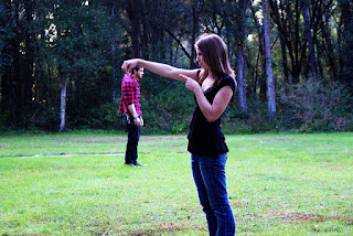
Trick photography apart, my intention is to draw your attention to the utmost importance one needs to attribute to the perception of the third dimension....THE DEPTH OF FIELD ....to get an idea of the relative distance of objects from one another...In ENT (as anywhere in life) and more so in an accurate ear examination and to come to correct diagnosis,Sterioscopic or Binocular vision is A MUST. To ensure this, the classical or conventional ENT Setup of Bull's Eye Lamp and Head Mirror is extremely useful... 


Wednesday, August 10, 2016

Dear Friends,
Trying something different to ensure that the vdos are compatible to run on mobile devices. Please bear with us.
A small vdo of ear cleaning in the OPD.
https://youtu.be/x4h4Vcs8PsI
Please peruse and give your feedback.
Amol.

Sunday, July 17, 2016

Dear Friends,
Adding one image and a commensurate vdo.
Please peruse.

Ear Examination with
Speculum

Also note 3 subtle points -
1) An attempt ALWAYS to keep the hand/s out of path of the light to ensure that no shadows are thrown inadvertently compromising the illumination of the area of interest.
2) Using the largest possible ear speculum with the objective of trying to examine the Tympanic Membrane in its entirety.
3) Introduction of the ear speculum is done in a gentle screwing action to prevent undue stimulation and avoid a possible vasovagal syncope.
Dear All
Uploading a small vdo clip of Ear Examination which adheres to the basic prerequisite of -
Consistent and Concentrated source of illumination in the form of the conventional setup of Bull's Eyelamp and Head Mirror.
The vdo also extolls on the virtue of -
1) First honest attempt for a naked eye examination (i.e. without Magnification)
2) Without instrumentation.


Sunday, August 10, 2014

Dear Friends,
It has been quite some time that I have posted anything on the blog.I say "quite some time" because 2  batches have already completed their clinical postings in ENT and a good amount of clinical taching has been accomplished (of course A BIG THANKS to your sense of commitment)...
Now posting a VDO of Acute Suppurative Otitis Media at the stage of perforation. Would want you to basically   to observe 2 things..
1) The classical "LIGHT HOUSE SIGN" .
2) The (?) therapeutic use of the Siegle's Pneumatic Speculum ...of sucking away the secretions.
Please peruse.....
Also do give me your feedback which acts like an elixir for my enthusiasm.
Till then....ADIEU
Amol.

Wednesday, January 29, 2014

Dear Aditya & ,
Uploading a VDO of Antrochaonal Polyp.You can see in the vdo that it is arising from right side (AC Poylps  are UNILATERAL) and as is its tendency to grow posteriorly towards the nasopharynx.This has essentially 2 effects - 1) They are not are not seen easily on routine anteior rhoinoscopy
               2) When they reach the nasopharynx,they cause a BILATERAL nasal blockage even when they are UNILATERAL.
Also PROBING is used to differentiate it from 1) Hypertrophied Turbinate which is
a) Sensitive to touch

b) Bleeds to touch
c) Hard in consistency
d) It cannot be probed all around as it is laterally attached.
Please watch carefully.
Regards,
Amol.