Tuesday, April 23, 2019

Sunday, April 21, 2019
Clinic @10
OTOSCOPIC SIEGELIZATION

As discussed in the previous post,most of the Otoscopes nowadays have a Siegel's attachment
USES of Siegelization
(A) DIAGNOSTIC
1) Magnification..2X
2) To assess the Mobility of the TM
3) To perform FISTULA TEST
(B) THERAPEUTIC
1) To perform aural toilet
2) To insufflate medication..usually Antibiotic powder
3) To break the synechiae between TM and Promontory in the early stages.
Clinic @9
EAR EXAMINATION WITH AN OTOSCOPE

When examining with an Otoscope, one may note
1) The Otoscope should be held like a pen.
2) The Otoscope should be held in the hand which side the ear is to be examined i.e. RIGHT for RIGHT and LEFT for LEFT.
3) While examining, the Ring finger and the Little finger of the hand holding the scope should rest on the cheek of the patient to ensure that there is NO inadvertent trauma ( to the Ear).
4) The other hand pulls the pinna to ensure straightening of the pinna.
ADVANTAGE/S
1)Self contained source of illumination..
2) Magnification ..2X (some newer models have more.
3) Facility for Siegeization.
DISADVANTAGE/S
1)Runs on dry battery cells..hence illumination over a period of time will decrease.
2) Uniocular vision....hence compromise of DEPTH OF FIELD.
Clinic @8
EAR EXAMINATION WITH SPECULUM

When one examines WITH a speculum,it may be noted
1) One should use the largest possible speculum so that the TM may be seen in one single position.
2) The speculum should be introduced in a gentle screwing movement so that there is NO VASOVAGAL STIMULATION...
Clinic @7
 EAR EXAMINATION WITHOUT SPECULUM
It may be noted
1) The use of right hand here to pull the pinna is WRONG...it could have potentially obstructed the path of light.
2) The direction in which the pinna is pulled.....UPWARD,OUTWARD and BACKWARD ensures that the EAC is straightened for satisfactory examination of the TM.
Clinic @6

Certain pertinent points to discuss taking benefit of the above image
1) The OPD setup ( Bull's Eye Lamp and Head Mirror)should be such that the source of illumination (the Bull's Eye Lamp here) and the reflector (the head mirror here) should be close to each other.......so with a RIGHT HANDED examiner, the mirror should be on the right eye of the examiner...for subtle adjustment/s of the mirror can be made with the dominant hand and accordingly the Bull's lamp should also be on the right side of the EXAMINER....behind the LEFT shoulder of the patient....one needs to remember here that the orientation of the patient and the examiner are opposite i.e. the EXAMINER'S RIGHT is the PATIENT'S LEFT and vice versa..
2) Initially, the examiner should make a honest and fervent attempt to examine WITHOUT instrument/s and WITHOUT magnification..
3) While performing examination, care has to be taken that the examiners' hand/s or intrument/s SHOULD NOT obstruct the path of illumination and vision.
P.S. Admit this post is too verbose....henceforth will try to be brief .
Clinic @5
EXAMINATION...
How?


How?

Conventional OPD Examination setup encompasses
1) Bull's Eye Lamp
2) Head Mirror
This setup ensures
a) CONSISTENT and CONCENTRATED illumination.
b) BINOCULAR vision.
Binocular vision is EXTREMELY MANDATORY for perception of DEPTH OF FIELD, the significance of which can't be underestimated after per viewing the following images...


Clinic @4
HISTORY TAKING..
Why..???
1) To come to a diagnosis...e.g. CSOM safe/unsafe
2) To know the etiology...e.g. Tonsilloadenoiditis Rhinosinusitis...as a cause for CSOM
3) To ascertain the severity of the condition...e.g. Mild/Moderate/Severe hearing loss...conductive/sensorineural
4) To identify any ( Impending) complication/s...e.g. Facial palsy, Gradenigo's Syndrome,Otogenic Meningitis
5) To know any co morbidities...e.g. DM, HT,IHD.
LEADING QUESTION.. a question which is framed in such a manner the answer to can be ONLY YES or NO...
Why..???
1) To come to a diagnosis...e.g. CSOM safe/unsafe
2) To know the etiology...e.g. Tonsilloadenoiditis Rhinosinusitis...as a cause for CSOM
3) To ascertain the severity of the condition...e.g. Mild/Moderate/Severe hearing loss...conductive/sensorineural
4) To identify any ( Impending) complication/s...e.g. Facial palsy, Gradenigo's Syndrome,Otogenic Meningitis
5) To know any co morbidities...e.g. DM, HT,IHD.
LEADING QUESTION.. a question which is framed in such a manner the answer to can be ONLY YES or NO...
Clinic @3
HISTORY TAKING...
The time alloted for history taking and examination for a long case in the MUHS University is 30 minutes.
It is expected of the candidate to present the history in a definite sequence...
1) Particulars of the Patient
2) Chief Complaint/s...to be ENUMERATED in the patients words verbatim in CHRONOLOGICAL order.
3) Onset,duration and progress
4) Past history
5) Personal history
6) Family history
7) \Drug history
8) H/o Allergy
9) Immunization history
10) History of Surgery in the past
11) GENERAL EXAMINATION
12) Local Examination..
Ears
Nose
Throat
Neck
13) Tuning Fork Tests...for ear case.
 Would like to humbly urge all as I have often quoted to follow the example of this marvelous feat of human engineering...THE PYRAMID...let the base of my history taking be so exhaustive that the pinnacle of my diagnosis will NEVER topple over....
The time alloted for history taking and examination for a long case in the MUHS University is 30 minutes.
It is expected of the candidate to present the history in a definite sequence...
1) Particulars of the Patient
2) Chief Complaint/s...to be ENUMERATED in the patients words verbatim in CHRONOLOGICAL order.
3) Onset,duration and progress
4) Past history
5) Personal history
6) Family history
7) \Drug history
8) H/o Allergy
9) Immunization history
10) History of Surgery in the past
11) GENERAL EXAMINATION
12) Local Examination..
Ears
Nose
Throat
Neck
13) Tuning Fork Tests...for ear case.

Clinic @ 2
Dear All,
THE MARKING SYSTEM..
1) Long case...20 marks
2) Short case...10 marks
3) Table...Viva Voce...10 marks
Instruments...03
Specimen...03
X Rays...02
Audiograms...02
LONG CASE..
Usually it is a Ear Case.. a case of CSOM safe
History taking..05 marks.
Examination..05 marks.
Diagnosis..05 marks.
Treatment..05 marks. (*)
(*) Treatment basically implies investigations and treatment.
THE MARKING SYSTEM..
1) Long case...20 marks
2) Short case...10 marks
3) Table...Viva Voce...10 marks
Instruments...03
Specimen...03
X Rays...02
Audiograms...02
LONG CASE..
Usually it is a Ear Case.. a case of CSOM safe
History taking..05 marks.
Examination..05 marks.
Diagnosis..05 marks.
Treatment..05 marks. (*)
(*) Treatment basically implies investigations and treatment.
Clinic @1
Dear All,
In pursuance of the clinical case discussion we had the other day, would like to share certain things,
THE UNWRITTEN PROTOCOL...
1) During the final MUHS practical examination, be appropriately dressed for the occasion.
2) After entering the examination hall, wish the examiner/s .
3) Be seated ONLY after being asked to do so.
4) AVOID use of short forms (acronyms) e.g. CSOM, ESR etc.
5) When the candidate is asked to explain any examination e.g. indirect laryngoscopy or any test e.g. tuning fork test, it is appropriate to preface the answer by saying " I WILL EXPLAIN THE PROCEDURE/TEST TO THE PATIENT IN A LANGUAGE WHICH HE OR SHE UNDERSTANDS".....creates a good impression on the examiner..
In pursuance of the clinical case discussion we had the other day, would like to share certain things,
THE UNWRITTEN PROTOCOL...
1) During the final MUHS practical examination, be appropriately dressed for the occasion.
2) After entering the examination hall, wish the examiner/s .
3) Be seated ONLY after being asked to do so.
4) AVOID use of short forms (acronyms) e.g. CSOM, ESR etc.
5) When the candidate is asked to explain any examination e.g. indirect laryngoscopy or any test e.g. tuning fork test, it is appropriate to preface the answer by saying " I WILL EXPLAIN THE PROCEDURE/TEST TO THE PATIENT IN A LANGUAGE WHICH HE OR SHE UNDERSTANDS".....creates a good impression on the examiner..
Monday, January 7, 2019
TUNING FORK TESTS
Tuning fork tests are an integral part of ear examination which help us assess the FUNCTION of the ear....HEARING.
There are many Tuning fork tests but the commonly performed are 3
1) Rinne's Test
2) Weber's Test
3) Absolute Bone Conduction Test
The tests essentially tell us whether the hearing loss is....
a) CONDUCTIVE...the pathology is in the EXTERNAL EAR +/- MIDDLE EAR.....Treatment is Medical +/- Surgical.
b) SENSORI NEURAL...the pathology is in the INNER EAR or beyond....Providing a commensurate hearing aid is the only treatment.
c) MIXED
So the competence to perform these tests properly is mandatory .
The tests need to be discussed under the following headings
Principle
Procedure
Observstion/s
Inference
Exception/s
RINNE'S TEST


There are many Tuning fork tests but the commonly performed are 3
1) Rinne's Test
2) Weber's Test
3) Absolute Bone Conduction Test
The tests essentially tell us whether the hearing loss is....
a) CONDUCTIVE...the pathology is in the EXTERNAL EAR +/- MIDDLE EAR.....Treatment is Medical +/- Surgical.
b) SENSORI NEURAL...the pathology is in the INNER EAR or beyond....Providing a commensurate hearing aid is the only treatment.
c) MIXED
So the competence to perform these tests properly is mandatory .
The tests need to be discussed under the following headings
Principle
Procedure
Observstion/s
Inference
Exception/s
RINNE'S TEST


ABSOLUTE BONE CONDUCTION TEST


Monday, June 5, 2017
Dear All,
Posting a few images of ears for clinical discussion.
Image No. 1)

Image No. 2)

Points to notice -
1) Both the images are of the RIGHT ear as is our protocol ( for matters of convenience).
2) Image No. 1 shows a big DEFECT in the Tympanic Membrane.
3) Image No. 2 shows multiple discrete chalky flakes on the Tympanic Membrane....these are typical TYMPANOSCLEROTIC patches which suggest old affection of the Middle Ear (probably an old c/o Otitis Media)...this is an extension of our discussion yesterday in the theory class on Otosclerosis.....this condition can mimic Otosclerosis (Differential diagnosis) in the sense that here too patient may complain of hard of hearing WITHOUT any h/o earache, otorrhoea ....the ONLY thing that will distinguish it from Otosclerosis is the ABNORMAL appearance of TM.
Points to ponder -
1) Which type of perforation does the Image No. 1 denote?
2) What is the type of CSOM here?
3) What are the Synonyms for this type of CSOM?
4) Waht is the Management of this condition?
Posting a few images of ears for clinical discussion.
Image No. 1)

Image No. 2)

Points to notice -
1) Both the images are of the RIGHT ear as is our protocol ( for matters of convenience).
2) Image No. 1 shows a big DEFECT in the Tympanic Membrane.
3) Image No. 2 shows multiple discrete chalky flakes on the Tympanic Membrane....these are typical TYMPANOSCLEROTIC patches which suggest old affection of the Middle Ear (probably an old c/o Otitis Media)...this is an extension of our discussion yesterday in the theory class on Otosclerosis.....this condition can mimic Otosclerosis (Differential diagnosis) in the sense that here too patient may complain of hard of hearing WITHOUT any h/o earache, otorrhoea ....the ONLY thing that will distinguish it from Otosclerosis is the ABNORMAL appearance of TM.
Points to ponder -
1) Which type of perforation does the Image No. 1 denote?
2) What is the type of CSOM here?
3) What are the Synonyms for this type of CSOM?
4) Waht is the Management of this condition?
Sunday, January 22, 2017
Dear Friends,
Posting some images


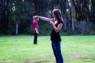


Posting some images


Trick photography apart, my intention is to draw your attention to the utmost importance one needs to attribute to the perception of the third dimension....THE DEPTH OF FIELD ....to get an idea of the relative distance of objects from one another...In ENT (as anywhere in life) and more so in an accurate ear examination and to come to correct diagnosis,Sterioscopic or Binocular vision is A MUST. To ensure this, the classical or conventional ENT Setup of Bull's Eye Lamp and Head Mirror is extremely useful...


Wednesday, August 10, 2016
Monday, July 18, 2016
Sunday, July 17, 2016
Dear Friends,
Adding one image and a commensurate vdo.
Please peruse.
Also note 3 subtle points -
1) An attempt ALWAYS to keep the hand/s out of path of the light to ensure that no shadows are thrown inadvertently compromising the illumination of the area of interest.
2) Using the largest possible ear speculum with the objective of trying to examine the Tympanic Membrane in its entirety.
3) Introduction of the ear speculum is done in a gentle screwing action to prevent undue stimulation and avoid a possible vasovagal syncope.
Adding one image and a commensurate vdo.
Please peruse.
 |
| Ear Examination with Speculum |
Also note 3 subtle points -
1) An attempt ALWAYS to keep the hand/s out of path of the light to ensure that no shadows are thrown inadvertently compromising the illumination of the area of interest.
2) Using the largest possible ear speculum with the objective of trying to examine the Tympanic Membrane in its entirety.
3) Introduction of the ear speculum is done in a gentle screwing action to prevent undue stimulation and avoid a possible vasovagal syncope.
Dear All
Uploading a small vdo clip of Ear Examination which adheres to the basic prerequisite of -
Consistent and Concentrated source of illumination in the form of the conventional setup of Bull's Eyelamp and Head Mirror.
The vdo also extolls on the virtue of -
1) First honest attempt for a naked eye examination (i.e. without Magnification)
2) Without instrumentation.
Uploading a small vdo clip of Ear Examination which adheres to the basic prerequisite of -
Consistent and Concentrated source of illumination in the form of the conventional setup of Bull's Eyelamp and Head Mirror.
The vdo also extolls on the virtue of -
1) First honest attempt for a naked eye examination (i.e. without Magnification)
2) Without instrumentation.
Sunday, August 10, 2014
Dear Friends,
It has been quite some time that I have posted anything on the blog.I say "quite some time" because 2 batches have already completed their clinical postings in ENT and a good amount of clinical taching has been accomplished (of course A BIG THANKS to your sense of commitment)...
Now posting a VDO of Acute Suppurative Otitis Media at the stage of perforation. Would want you to basically to observe 2 things..
1) The classical "LIGHT HOUSE SIGN" .
2) The (?) therapeutic use of the Siegle's Pneumatic Speculum ...of sucking away the secretions.
Please peruse.....
Also do give me your feedback which acts like an elixir for my enthusiasm.
Till then....ADIEU
Amol.
It has been quite some time that I have posted anything on the blog.I say "quite some time" because 2 batches have already completed their clinical postings in ENT and a good amount of clinical taching has been accomplished (of course A BIG THANKS to your sense of commitment)...
Now posting a VDO of Acute Suppurative Otitis Media at the stage of perforation. Would want you to basically to observe 2 things..
1) The classical "LIGHT HOUSE SIGN" .
2) The (?) therapeutic use of the Siegle's Pneumatic Speculum ...of sucking away the secretions.
Please peruse.....
Till then....ADIEU
Amol.
Wednesday, January 29, 2014
Dear Aditya & ,
Uploading a VDO of Antrochaonal Polyp.You can see in the vdo that it is arising from right side (AC Poylps are UNILATERAL) and as is its tendency to grow posteriorly towards the nasopharynx.This has essentially 2 effects - 1) They are not are not seen easily on routine anteior rhoinoscopy
2) When they reach the nasopharynx,they cause a BILATERAL nasal blockage even when they are UNILATERAL.
Also PROBING is used to differentiate it from 1) Hypertrophied Turbinate which is
a) Sensitive to touch
b) Bleeds to touch
c) Hard in consistency
d) It cannot be probed all around as it is laterally attached.
Please watch carefully.
Regards,
Amol.
Uploading a VDO of Antrochaonal Polyp.You can see in the vdo that it is arising from right side (AC Poylps are UNILATERAL) and as is its tendency to grow posteriorly towards the nasopharynx.This has essentially 2 effects - 1) They are not are not seen easily on routine anteior rhoinoscopy
2) When they reach the nasopharynx,they cause a BILATERAL nasal blockage even when they are UNILATERAL.
Also PROBING is used to differentiate it from 1) Hypertrophied Turbinate which is
a) Sensitive to touch
b) Bleeds to touch
c) Hard in consistency
d) It cannot be probed all around as it is laterally attached.
Please watch carefully.
Regards,
Subscribe to:
Posts (Atom)